Generally, no prior preparation, like fasting or sedation, is required. Your health care provider will explain the procedure to you and offer you the opportunity to ask questions that you might have about the procedure. Be sure to discuss any concerns with your health care provider prior to the procedure. There may be other risks depending on your specific medical condition. If it is necessary for you to have a spinal X-ray, special precautions will be made to minimize the radiation exposure to the fetus. Radiation exposure during pregnancy may lead to birth defects. If you are pregnant or suspect that you may be pregnant, you should notify your health care provider. Risks associated with radiation exposure may be related to the cumulative number of X-ray exams and/or treatments over a long period of time. It is a good idea to keep a record of your past history of radiation exposure, like previous scans and other types of X-rays, so that you can inform your health care provider. You may want to ask your health care provider about the amount of radiation used during the procedure and the risks related to your particular situation. The spinal cord carries sense and movement signals to and from the brain and controls many reflexes. The spinal cord is surrounded by the bones of the spine and a sac containing cerebrospinal fluid. The spinal cord, a major part of the central nervous system, is located in the vertebral canal and reaches from the base of the skull to the upper part of the lower back. The 4 coccygeal vertebrae fuse to form 1 bone, called the coccyx or tailbone. The lumbar area consists of 5 vertebrae in the lower back. The thoracic area consists of 12 vertebrae in the chest. The cervical area consists of 7 vertebrae in the neck.

The spinal column is made up of 33 vertebrae that are separated by spongy disks and classified into distinct areas: Please see these procedures for additional information.

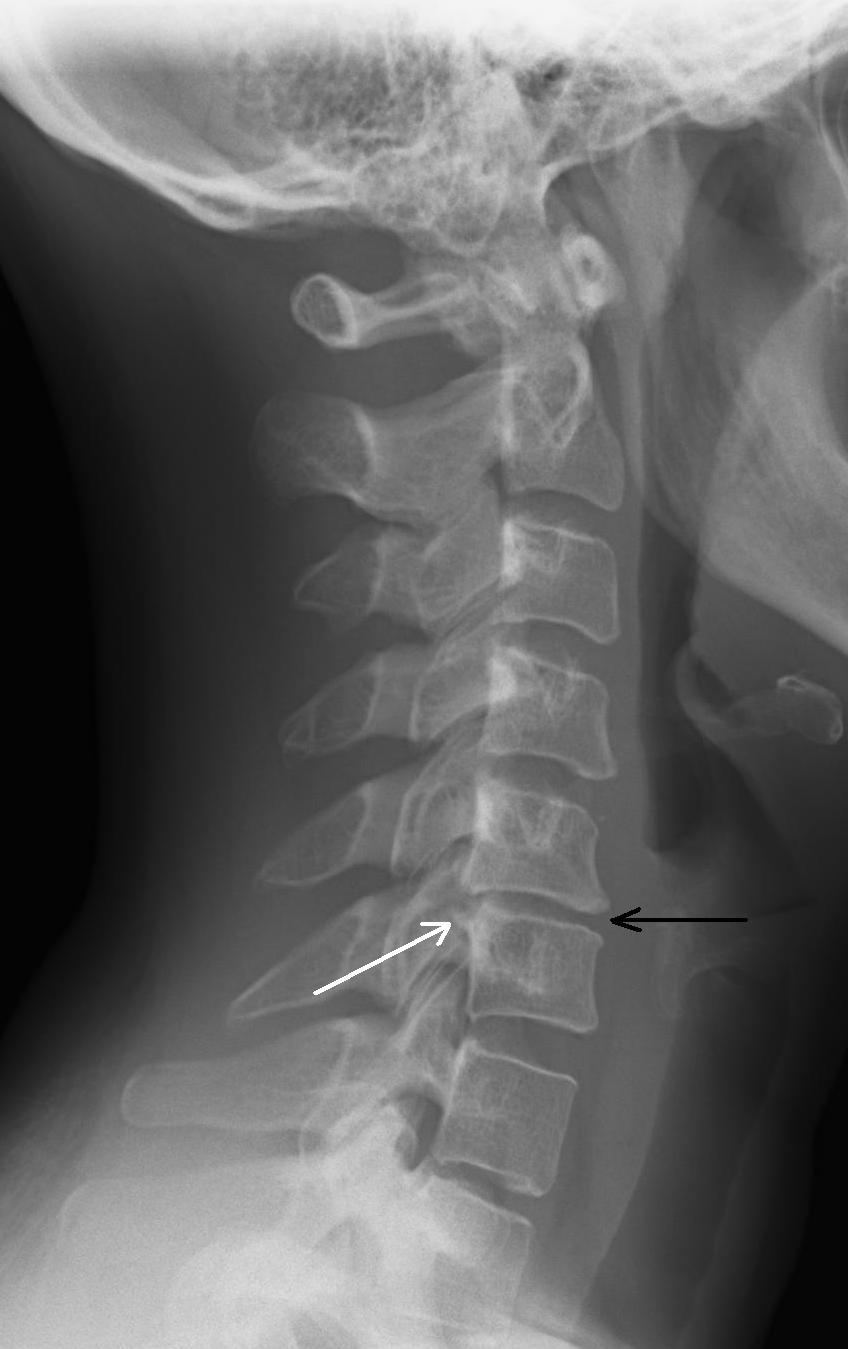
Other related procedures that may be used to diagnose spine, back, or neck problems include myelography (myelogram), computed tomography (CT scan), magnetic resonance imaging (MRI), or bone scans. X-rays of the spine may be performed to evaluate any area of the spine (cervical, thoracic, lumbar, sacral, or coccygeal). It appears as a dark line in the white bone. At a break in a bone, the X-ray beam passes through the broken area. A bone or a tumor, which is denser than soft tissue, allows few of the X-rays to pass through and appears white on the X-ray. The soft tissues in the body (like blood, skin, fat, and muscle) allow most of the X-ray to pass through and appear dark gray on the film. It depends on the amount of X-rays that penetrate the tissues. Images are made in degrees of light and dark. When the body undergoes X-rays, different parts of the body allow varying amounts of the X-ray beams to pass through. Instead of film, X-rays are now typically made by using computers and digital media. X-rays pass through body tissues onto specially-treated plates (similar to camera film) and a "negative" type picture is made (the more solid a structure is, the whiter it appears on the film). X-rays are made by using external radiation to produce images of the body, its organs, and other internal structures for diagnostic purposes. These include diagnosing tumors or bone injuries. Standard X-rays are performed for many reasons. X-rays use invisible electromagnetic energy beams to make images of internal tissues, bones, and organs on film. What are X-rays of the spine, neck or back?


 0 kommentar(er)
